
It's Just a Strain - Kurt Jepson
By Kurt Jepson

Summer is under way and as such your training plans likely include elevated intensity and frequency of hard efforts. You may have some races scheduled on foot or two wheels. This time of year, your exposure for isolated muscle injury increases. As seemingly benign as a muscle “pull” appears, they can snowball into a recalcitrant, recurrent problem. A simple “strain” can at the least limit timely progression in training, and if mismanaged, sideline the athlete for weeks or months.
This post will discuss what a muscle “pull” is and how to best avoid complications.
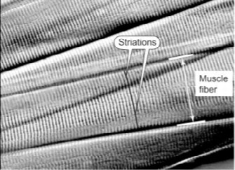
Skeletal muscle fibers are composed of large cells of up to 100 um in diameter and many centimeters long in some groups (Feher J. Quantitative Human Physiology, 2nd edi. 2017). The cells are “multinucleated” which, due to their length, allows protein synthesis and regeneration from many sites. Under high magnification the cells present with a “banded” appearance and have “striations” which run perpendicular to the length of the muscle itself. This organization forms the basis of overlap and shortening of the unit during contraction.
Perpendicular A- and I-bands (source, J Feher).
Longitudinally muscle cells are organized into “myofibrils” which are in turn composed of many “filaments”. Filaments may be thin (actin) or thick (myosin) and overlap during band contraction. Cytoskeletal proteins are the structural building block of muscle cells and have the very important role of transmitting force throughout the cell (Lieber R, Shah S, Friden J. Cytoskeletal disruption after eccentric contraction induced muscle injury. Clin Orthop Relat Res. 2002). Other construct of the cells include; sarcoplasmic reticulum, mitochondria (the energy source of cells), and glycogen granules.
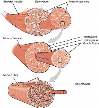
From a macroscopic to microscopic view, skeletal muscle transitions from fascicles to fibers to filaments. All are surrounded by cellular matrix material as depicted below.
Like all tissue, muscle cells are in a constant state of change. James Tidball sums it up best; “Skeletal muscle continuously adapts to changes in its mechanical environment through modifications in gene expression and protein stability that affect its physiological function and mass. However, mechanical stresses commonly exceed the parameters that induce adaptations and instead produce acute injury” (Mechanisms of muscle injury, repair, and regeneration. Compr Physiol. 2011; 1(4)).
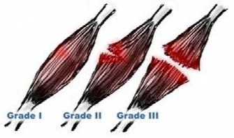
Muscle strain injuries are clinically classified based on the degree of fiber involvement and degradation. Grade I strain`s equate to microscopic “tearing” of the cells, grade II is represented by partial structural compromise and a grade III lesion involves complete disassociation of the muscle unit. A grade II strain encompasses the largest severity spectrum, as fiber involvement may be +/- 10% or +/- 80%. Resultant dysfunction and time to resolution, need for surgical intervention and/or required immobilization correlates to the grade of injury.
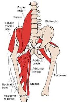
Some muscle groups are more predisposed to injury than others based on composition and function. Fast twitch type II dominated muscles are more prone to injury than other types. Also, muscles which act on two joints and have triplanar biomechanical demands such as the hip adductors (groin) are at enhanced risk of injury (Neumann D. Kinesiology of the hip: A focus on function. JOSPT; 2010). Hamstrings, hip flexors, paraspinals and the calf complex also fall into this category.
Other specific risk factors have been identified such as; increasing age (Orchard 2001, Verral et al 2001, Arnason et al 2008), having sustained a previous strain (Seward et al 1993, Emery et al 2001) and muscle weakness relative to the antagonist group or interestingly, the contralateral side (Tyler 2001, Orchard et al 1997).
When a muscle succumbs to Tidball`s quote above, it is usually the result of contractile forces exceeding fiber tensile integrity via a decelerating or eccentric contraction of the muscle group. Examples include the hamstrings as they decelerate the lower limb (knee extension, hip flexion) while sprinting, or while they control the forward progression of our core/hips during late stage, high velocity, double pole.
There is evidence to support that this mechanism of injury occurs most frequently when the muscle is weakened by elongation (minimal band overlap at the fibril level). This concept will be considered when we discuss rehabilitative exercise.
Less frequently, a strain may occur as a result of excessive elongation load at its end range (aggressive stretching), or via a vigorous concentric shortening (max lifting).
Injury at the cellular level includes mechanically induced disruption of the myofibrils, the membranes (-mysiums), and/or sarcolemmal. The myofibril protein Desmin, has been shown to be particularly important as a force transmitter/dispersion compound to the muscle tendon junction as well as the surface of the muscle itself. Animal model studies show this protein is rapidly lost following high intensity exercise or injury (Lieber et al, Cytoskeletal disruption after eccentric contraction induced muscle injury. Clin Orthop Relat Res. 2002). Disruption of the cell membrane allows unregulated influx of calcium, inflammatory cells and activation of enzymes that promote and regulate repair.
Although this myogenic sequence of events post- acute injury is very efficient in modulating repair, it is lacking or dysregulated in chronic muscle injuries. Hence, the “stubborn” nature of some recurrent strain injuries if mismanaged initially. Chronic strains require repetitive mechanical loading to promote a quality repair.
Not all muscle groups respond to injury the same. Fiber type (I vs II vs hybrids), capillary density, neural supply, and individual morphology all play a role in the healing timeframe of muscle cells. In general, fast twitch type II dominated muscles take longer to normalize and undergo repair (i.e. hamstrings).
Management of strains follows an algorithm similar to other soft tissue injuries.
First, recognize the issue and adjust the training load. There are very few muscle injuries an athlete can successfully “work through”. If it`s significant enough to affect a normal movement pattern, even transiently, it`s significant enough to treat and adapt training.
Second, utilize any anti-inflammatory tools you`ve had success with in the past to allow early therapeutic movement. Use them sparingly. Recall that the cascade of inflammatory events at the cellular level is the bodies` mechanism to promote repair. It is unlikely the athlete can significantly override these events to the point of adversely affecting the end outcome by employing anti-inflammatory modalities early on. That said, I tell my patients if the discomfort is significant enough to interrupt your normal sleep pattern, then it is worthy of addressing in the short term with OTC medicines (approved by your physician), icing, electrical modalities, etc. A review of a post on this site entitled; “Icing after injury; evidence based or “folklore”?”, discusses this topic in more depth.
Third, prepare the tissue for the therapeutic loads to come. By that I`m referring to employing gentle mechanical stimulus to the involved area. Low load, repetitive motion stimulates capillary/lymphatic exchange, bed proliferation and sensory re-biasing. Light activity in a pool, on a stationary bike or gentle manual soft tissue techniques are useful. Do not “foam roll” an acute muscle strain, particularly if the injury mechanism involved compression/contusion. A “bruise” injury typically involves the sarcolemma or membrane components of the muscle and compressive rolling further traumatizes these structures.

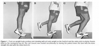
The key is keeping the initial work load small. This is not a new concept. In the late 1800`s a French surgeon commented on his view of exercise in the early stages of injury convalescence. Just Lucas-Championniere eloquently said; “Any movement, so long as it is not injurious by means of it`s amplitude, promotes repair.” That sentiment is often lost in modern medicine where prescribed “rest” of an injured region is to restrictive.
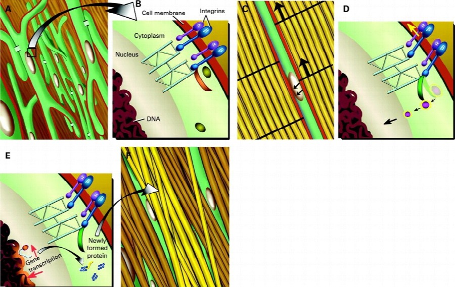
This brings us to the fourth principle of muscle strain management, “mechanotransduction” (McElhany et al, J Biomech,1968 (1)).
In short, mechanotransduction refers to the process where mechanical loading results in a cellular response. Cellular responses collectively result in structural change and compensation. Wolff`s Law is a similar concept related to the physiologic response of bone tissue to stressors. Mechanotransduction is broken down into three steps per Khan and Scott (Br J Sports Med.2009); mechanocoupling, cell to cell communication and the effector response. Let`s briefly define these steps of tissue adaptation.
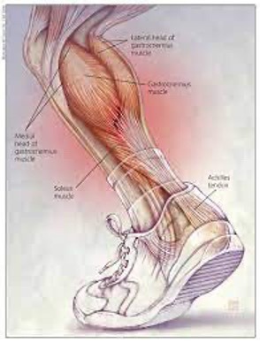
Mechanocoupling refers to the imparted load to a tissue in the form of shear or compression. An example would be the forces an Achilles Tendon experiences during a run. There are both compressive loads as it rounds the corner of the calcaneus to its insertion, as well as inline tensioning and shear via ground reaction forces and calf muscle contraction. These physical “perturbations” (repetitive load challenges) signal the cells to undergo a variety of chemical changes within and among cells in order to maintain structural integrity locally.
Cell to cell communication involves a wide spread chemically mediated adaptation along the entire muscle tendon unit. Calcium and Inosital Triphosphate are components of signaling proteins within the extracellular matrix that initiate change in “location 2“ via “location 1” mechanical stimulus (Khan and Scott), despite location 2 never experiencing mechanocoupling directly.
Effector cell response focuses on protein synthesis at the extracelluar matrix boundary stimulated by mechanical load. This detailed process is well understood for bone regeneration but less so for muscle tissue. In short, each connective tissue in the body is thought to have a “set point” and if stress is taken away, the tissue weakens due to a lack of “signal” to retain structure. If stress is added, the tissue adapts via protein synthesis and hypertrophy. In muscle, the growth response to “overload” is mediated by Mechanical Growth Factor (MGF) and a form of Insulin-like Growth Factor (IGF-I) (Goldspink G. Gene expression in muscle in response to exercise. J Muscle Cell Motil; 2003: 24). The resultant stimulation of satellite cells via MGF leads to hypertrophy if the load was tolerated, or “scar” production if the mechanocoupling force was extensive enough to cause injury. Muscle scar lacks the elastic, tensile and energy storage characteristics required for sound function.
So how does the science translate into muscle strain management? Below are some basic guidelines;
Phase 1; immediate post insult (day 1-7):
Recognize the severity/injury grade and adapt training. Grade 1 and most grade 2 strains are typically managed conservatively. Grade 3 lesions require medical intervention. Seek diagnosis as appropriate. Limited use of ice/ OTC anti-inflammatory meds, limited immobilization based on severity and “relative” rest. If it`s a skiing induced injury, bike. If it`s a running induced injury, DP, etc. L1 work can continue but may need to be abbreviated/adapted.
Phase 2; sub-acute/early healing stage (day 8-14):
Mechanocoupling may have already begun via adapted training above (ie stationary cycling). If not, low “amplitude” load may be initiated. This can be in the form of mid-range muscle/tendon specific submaximal isometrics (10-20% volitional efforts, 60-90 sec each x 4 reps, daily), antagonist activation (i.e. slow controlled quad work for a hamstring pull) or gentle manual therapy if NOT originally a compressive/contusion injury. The athlete should focus on gym activities that “work away” from the injury site. For instance, if the athlete has experienced a hip flexor strain, she/he should focus on controlled gluteal and low back exercises. If it`s a triceps “pull”, work the elbow flexors, etc.
NO stretching!! It is very difficult to place tensile load along a muscle injury site and not exceed the mechanocoupling load that signals repair, versus that which further degrades the myofibrils and associated membranes. If you perceive a stretch, you`ve already gone too far and the injury regresses back to the acute inflammatory stage.
Phase 3; early remodeling stage (day 15-30):
Mechanocoupling loads may be advanced in mode and resistance level. Review a prior NTS post on, “The Role of Eccentric Exercise in Early Season Training” for guidance. In general exercises should progress from; isometric to eccentric to concentric to weight bearing closed chain activities to agility/ balance work and lastly, to heavy eccentric work. Progression is regulated via resultant discomfort as graded on a 0-10 scale. Advancement in exercise mode or load should not cause one`s pain to exceed 2 points above the resting value and/or fail to resolve back to baseline within 12 hours post exercise bout. Failure to do so dictates regression back to the prior exercise mode and dose.
Anti-inflammatory modalities likely have little positive input at this stage and may be discontinued.
Phase 4; functional remodeling stage (31+ days):
Targeted rehabilitation exercise can now include more combined exercises/lifts (i.e. deadlifts, squats, cycling hill sprints, and early plyo- deceleration activities). No “explosive” concentric functional exercise (i.e. bounding) should be reintroduced until week 5 post injury at the earliest,
later if there is tendonitis as a secondary symptom or an extensive grade 2 lesion. Scar tissue may be adequately formed at the lesion site but has not had time to undergo advanced mechanotransduction, whereby taking on structural characteristics of the original muscle tissue.
Agility and balance work should be accelerated and shift to skiing specific drills. Tri-planar movement patterns are particularly important for 2 joint muscles such as the hip adductors and hamstrings. The contralateral limb and core should receive equal attention.
This is also the time to add heavy eccentric exercise to the rehab program. A classic “up with two down slow one” approach in the gym is vital to signaling tissue adaptation, tensile integrity, hypertrophy and functional contractile competence. Four to 6 sets of 4-6 reps at 80% MVC load is generally accepted as adequate stimulus for neural and tissue adaptations. Recall that most muscle strain injuries arise from an overload while in eccentric contractile mode. Do not skip this step! Twice a month eccentric work in the gym, indefinitely, will provide prophylactic input.
This also the appropriate timeframe for addition of static flexibility exercises back into her/his training. The debate on static stretching continues in the literature. McHugh and Cosgrave authored an excellent retrospective review of the literature in 2010 and I encourage athletes, coaches, and program designers to read it (To stretch or not to stretch: the role of stretching in injury prevention and performance. Scand J Med Sci Sports. 2010; 20: 169-181). One clinically relevant point focuses on the stretching induced shift of a muscle`s angle-torque relationship during contraction at end range. There is some evidence that end range stretching may provide protective input regarding strain injury.
As with any injury, associated timeframes with “time to cure”, tolerance for exercise activities and appropriate progression of rehab are physiologically specific to the athlete based on age, fitness, injury classification, prior injury, systemic health, and rehab compliance. Logically adjusting the athlete`s program chronologically is a given. A grade 1 strain may only need a week to get through the phases of rehab where as a significant grade 2 strain may take 2 months. Remember, you will rarely “work through” a muscle strain injury successfully. Taking short cuts often results in being haunted down the road by recurrence. How many times have you heard, “ya, I keep pulling my hamie”,……
Manage it right the first time. It may not be “just a strain”.
Enjoy the summer!