
SAFE AND SPECIFIC CORE CONDITIONING
SAFE AND SPECIFIC CORE CONDITIONING
By Kurt Jepson
We`ve spent the last few articles looking at hip and knee pathology related to various anatomic and kinematic inputs. Central to the corrective concept was the element of core stability. This seems like a good time to discuss safe and effective core conditioning, particularly for those athletes with a history of lumbar pain or those currently rehabilitating the same.
Few topics in the Sports Medicine and fitness literature over the last 15 years have garnished as much attention as “Core Conditioning”. Recent research has provided coaches, athletes and clinicians with valuable insight into muscle recruitment as well as recognizing potentially injurious inputs. “Activating” core musculature is not difficult. Most athletes are familiar with systems of exercise that effectively target core groups. The difficulty lies in weighing the relative “cost benefit” of performing readily accepted exercises.
The full content of this piece may be found at SkiTrax.com by searching my name.
To begin we must first review some basic anatomic and arthrokinematic characteristics of the human spine and pelvis.
The spine provides a structural foundation for attachment of ligaments and tendons, muscles, as well as an axis for core and hip movements.2Most importantly it provides bony protection for the spinal cord and it`s laterally projecting nerves. Each of the 24 plus vertebral bony segments is specific in its structural characteristics based on location, load bearing responsibilities, motion contributions and its relationship to neighboring extremities. For example, our Lumbar vertebrae, discs and ligaments are much larger and have a denser construct than their cervical counterparts because of the gravitational load on our trunk and exposure to powerful movements of our lower extremities. Each functional grouping (2+ vertebrae) of segments must balance movement, stability, and protection of neural structures contained within their boundaries. Your body considers your spinal cord to be quite vital, and would never encourage excessive vertebral movement or induced instability as this would likely endanger your nervous system. The lumbar spine is particularly sensitive to excessive rotatory inputs.
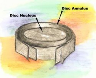
A key structure helping to maintain the balance between segmental movement and stability is the intervertebral disc. It may be compared to a jelly donut. It is composed of fibrous tissue peripherally called the annulus (donut), and has a viscous center (jelly) called the nucleus. Collectively it is composed of 80%+/- water based material depending on one`s age. Discs degrade and dehydrate with age, hence our relative loss in stature from our second to seventh decade. The disc is a primary connection between adjacent vertebral segments and as such functions as a ligament by connecting bone to bone. It must therefore have all required tensile strength and load bearing characteristics of ligament tissue, yet be subtle enough to allow mutliplaner movements of the axial spine. It accomplishes this feat through a complex system of annular fiber matrix design and nuclear hydraulics. The disc however is not perfect and can sustain focal or generalized failure zones, particularly within the annular ring (donut).3Cyclic loading in excess of its mechanical tolerances for spinal rotation and flexion can lead to annular wall compromise, nuclear bulging, frank herniations of the nucleus, fragmentation, and/or nuclear enzymatic leakage into adjacent space. The resultant irritation of nerve or soft tissue causes central or radicular pain, muscle spasm, and dysfunction.
Undampened trunk rotation is particularly stressful to lumbar structures, especially the annular tissue matrix. Structural fatigue and thus stability compromise of the disc can lead to “buckling” of the spine under load. The analogy of a stack of children`s building blocks arranged with characteristic spinal curves probably best exemplifies the relative instability of an unsupported vertebral column. It takes very little unopposed directional force to destabilize the blocks. Our discs, interspinous ligaments, and musculature all help oppose and negate directional forces encountered during sport and daily activities. Such structures surround the spine and when acting in concert provide what McGill refers to as “hoop forces”. 5 The analogy of a tent with balanced guy lines lending to the central structural support provides a good visual. . Research has repeatedly demonstrated that core strength and postural coordination aids in the dispersion of potentially injurious forces traveling inward toward the spine. 6 ,7, 8,
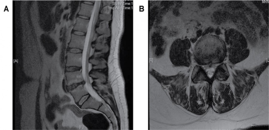
MRI of the lumbar spine with discs clearly visible. Note L4-5 discal degeneration.
So how does one define safe core load in the adult population? There will always be some variance based on individual genetics, age, anatomy and physiology, but we do have reliable science with which to establish reasonable load ranges. Various investigators have looked at spinal load to failure, measured in Newtons (N), in vivo and via cadaveric analysis.10, 11Others have analyzed the effect dynamic muscle action maintaining neutral spine has on load dispersion and tolerance. Position sense, balance and agility also have beneficial inputs to spinal load tolerance.
Cholewicki found that a cadaveric spine “buckled” with as little as 90N (~20lbs) of compressive force (think children`s blocks). 12He also found that as little as 10% maximum volitional contraction (MVC) of circumferential musculature added significant “stiffness” and stability to the spine in another study using live subjects. He also noted that competitive weight lifters quite readily handled loads of 2500N without compromising their neutral spinal position.
In other studies, Cripton 12 found sheer loads of 2000-2800N to be damaging to cadaver specimens, obviously void of working muscles. Adams, et al 10 found that in young men as little as 1200-1500N was injurious to the spine if there was no attention paid to posture. Gunning 8 noted that utilizing a neutral spine position during load testing increased his subject`s tolerance by 25-45%. Combining these and other works data into a numerical “safe zone” for spinal loading, we can target approximately 2500N and perhaps an additional 45%, if one is able to maintain a neutral ,stable spine throughout the exercise movement, as potentially safe loads.
So now let`s look at some in vivo spinal load data obtained by Axler, McGill, et al 13 recorded during common core and lower extremity exercises;
Straight leg full sit up- 3506N
Bent leg full sit up – 3350N
Quarter sit up- 2392N
Hanging bilateral straight leg raise- 2805N
Isometric side bridge- 2585N
It is important to note that the load to failure studies were examining peak load to failure. In the skiing population we need to be sensitive to cyclic tissue loading over time which usually entail lower loads delivered month after month, year after year.
Obviously not all high load postures can be avoided completely in the gym. We would however, like to select those exercises with high muscle recruitment and functionality if we are going to subject the spine to load. Skiers know the core groups they should target in the gym. Back extensors, deep spine rotators, ab`s, oblique’s, lateral hips, to name a few.
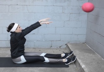
If we look at McGill`s work and compare two exercises used to target the abdominal groups, we see the trade off of high recruitment versus injurious spinal load over time. The straight knee full sit up activates the rectus abdominis (RA) to 121% of maximal volitional contraction (MVC). The exercise creates high activation but the cost to the spine significant at 3506N and over time would likely be injurious. The isometric side bridge by contrast recruits the RA to 48% MVC but at a very tolerable 2585N load to the spine. Yes, 48% is low in comparison but recall that as little as 10% MVC can add significant stability to a spine.
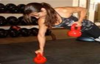
Below: High activation but at a high “cost” to lumbar structures.
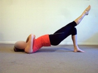
In 2007 Ekstrom et al 14 did an EMG analysis of more than 15 common core, hip and thigh muscle exercises and graded their relative value based on MVC recruitment abilities. Their findings incorporate nicely into this discussion as it applies to cross country skier gym programs. To summarize that study, back paraspinal /erector spinae recruitment was best achieved using, in order of efficiency; unilateral bridge with a straight leg raise (SLR), the side bridge, regular supine bridge, and the quadruped alternate leg/arm raise (Bird Dog) exercises. All had > 35% MVC and low cost to the spine provided subjects paid attention to neutral postures. Abdominal activation occurred via; side bridge, prone bridge/plank, Bird Dog and again the unilateral bridge with a concurrent SLR. Glut Medius activation occurred via ; side bridge at 74% MVC , unilateral bridge with SLR, lateral step ups, Bird Dogs and a close 5thwas the side lying leg raise. All of these muscle groups are essential to sound Nordic skiing technique. Including some of these exercises into a strength program would seem obvious.
Below: unilateral bridge with concurrent straight leg raise and plank row variation
Every athlete and coach should design programs with attention to individual needs, assessment of athletic competency and development, athlete goals, history of injury and mindful consideration of the potential “cost” of some exercises over the career of the athlete. Those athletes rehabilitating low back pain would likewise benefit from including some of these exercises into their therapeutic regime, directed by a clinician, particularly lumbar rotator isometric conditioning.
Kurt K Jepson, PT,SCS
REFERENCES:
- McGill SM. Low Back Exercises: Evidence for Improving Exercise Regimens. Phys Ther. 78, pp 754-765, 1998
- Magee DJ. Orthopedic Physical Assessment. Philadelphia: WB Saunders Company; p 467, 1992.
- Birrer R, Jepson KK. Low Back Pain: A Focused Approach. Consultant. July: 993-1035, 2003.
- Bogduk N. Pathology of Lumbar Disc Pain. J Manual Medicine. 5: 72-79, 1990.
- McGill SM. Low Back Stability: From Formal Description to Issues for Performance and Rehabilitation. Exer and Sport Sci Reviews. 29; 26-31, 2001.
- Parkhurst TM, Burnett CN. Injury and Proprioception in the Lower Back. J Ortho Sports Phys Ther. 19 (5); pp 282-295, 1994.
- Ekstrom RA, Osborn RW, et al. Surface Electromyographic Analysis of the Low Back Muscles During Rehabilitation Exercises. J Ortho Sports Phys Ther. 38; pp 736-745, 2008.
- Cholewicki J, Van Vliet JJ. Relative Contribution of Trunk Muscles to the Stability of the Lumbar Spine During Isometric Exertions. Clin Biomech. 17; pp 99-105, 2002.
- Cholewicki J, Greene HS , et al. Neuromuscular Function in Athletes Following Recovery From a Recent Acute Low Back Pain Injury. J Ortho Sports Phys Ther. 32; pp 568-575, 2002.
- Adams M, Dolan P. Recent Advances in Lumbar Spinal Mechanics and their Clinical Significance. Clin Biomech. 10 (3) p 3, 1995.
- Cholewicki J, McGill SM, et al. Lumbar Spine Loads during Lifting Extremely Heavy Weights. Med Sci Sports Exer. 23 (10); pp 1179-1186, 1991.
- Cripton P, Berlemen U, et al. Response of the Lumbar Spine due to Shear Loading. Injury Prevention Through Biomechanics. Detroit. Wayne State University. p 111, 1995.
- Axler CT, McGill SM . Low back Loads over a Variety of Abdominal Exercises: Searching for the Safest Abdominal Challenge. Med Sci Sport Exer. 29 (6), pp 804-810, 1997.
- Ekstrom RA, Donatelli RA , et al. Electromyographic Analysis of the Core Trunk,Hip, and Thigh Muscles During 9 Rehabilitation Exercises. J Ortho Sports Phys Ther. 37 (12) ; pp 754-762, 2007