
“YUP,… ROLLED IT!” (Lateral ankle sprains)
By Kurt Jepson
Phase 3, re-activation and strength, 2-4+ weeks

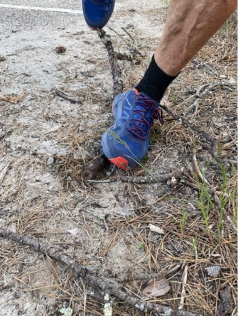
It`s going to happen at least once in your athletic career, if it hasn`t already, you are going to “roll” your ankle. It may result from stepping on a root trail running or getting snagged in a crack roller skiing, but it`s going to happen! What is an ankle sprain? How do you manage it to get back to training quickly?
Inversion sprains of the ankle are one of the most common lower extremity injuries associated with sporting activities. Injury to the lateral (outside) ligament complex comprise 85% of all ankle sprains (Gross MT, Phys Ther, 67, 1987) and up to 73% of individuals have residual symptoms including pain, repeat sprains and/or chronic instability which alters their activity level (Yeung et al, Br J Sports Med. 1994). Over 75% of lateral ankle sprains involve the anterolateral stabilizing complex/ligaments (Fong et al 2007, Roos et al 2017) in isolation with the remaining ~25% of sprains also involving the medial (Deltoid) ligaments and the more proximal Tibial-Fibular/Syndesmosis complex. Given it`s large size, thickness and multipennate design, the Deltoid ligament medially is rarely sprained without significant destabilizing input to the ankle.
A meta-analysis of 181 studies by Doherty et al in 2014, showed females experienced twice as many lateral ankle sprains as males. The incidence of injury decreased with age for both genders. A U.S. Military Health System 5-year cohort study of 30,910 patients demonstrated that 22.8% of ankle sprains involved an associated fracture (Rhon et al. IJSPT. 2021;16(5)). There is a saying in sports medicine regarding ankle injuries; “…if it`s locally and exquisitely tender, swollen and discolored, it`s broken until proven otherwise!”.
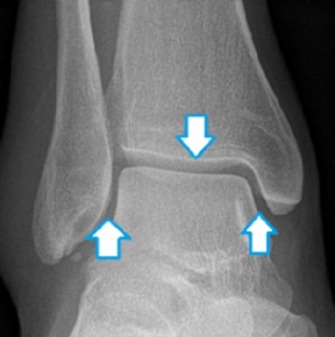
The bony anatomy of the ankle includes the distal tibia (medial malleolus) and fibula (lateral malleolus) which form the “mortise” superiorly. The superior aspect of the talus, referred to as the “dome” comprises the joint inferiorly. The talus is uniquely shaped in that it is relatively narrow posteriorly and widens anteriorly. As such, when the ankle is dorsiflexed (pulled upward), the wider anterior portion of the talus pivots and “wedges” into the mortise. This approximation limits excess motion and provides stability. Dorsiflexion is therefore a position of architectural stability through “joint partner” compression and congruency. Conversely, when the ankle/foot is pointed downward (plantar flexed), the ankle loses positional stability.
Ankle Mortise components below;
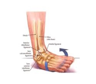
Arthrokinematically the ankle is often referred to as a “Hinge Joint” with motion occurring primarily in one plane (sagittal). In reality, ankle motion occurs in all three planes and there is obligatory inversion (in toe) as the ankle plantar flexes and obligatory eversion (out toe) as the ankle dorsiflexes. The “mortise joint” works in close concert with the sub-talar joint below. As such, the classic mechanism of injury to the anterolateral ligaments involves a combination of plantar flexion, inversion and supination when architectural stability is at a minimum.
The ankle joint is in part stabilized by surrounding ligaments (“static” stabilizers). Ligaments connect bone to bone, verses tendons which connect muscle to bone. Muscles extrinsic and proximal to the ankle joint encompass the front (anterior), rear (posterior), inside (medial) and outside (lateral) regions of the lower leg. When contracted, they provide significant “dynamic” stabilization via tensioning of their respective tendons which cross the ankle, inserting on the hind and midfoot. Residual joint stability occurs via bony approximation, joint compression, and neurokinetic pathways such as proprioception (joint position sense). Proprioception is a major contributor to balance and agility. McHugh and Tyler, et al saw a 77% reduction in ankle injuries in a population of “high risk” student athletes following the completion of a foam pad stability training program pre- and interseason (The Effectiveness of a Balance Training Intervention in Reducing the Incidence of Noncontact Ankle Sprains in High School Football Players, Amer J Sports Med. 35 (8) 2007).
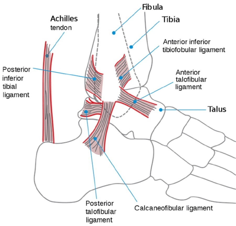
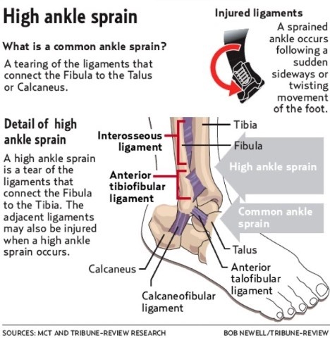
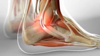
Anterolateral Ankle Ligaments;
For ease of discussion, each ligament is referred to via initials. For example, the ATF (Anterior Talo-Fibular) connects the anterior portion of the talus bone to the fibula, the CF (Calcaneofibular) spans the fibula and calcaneus, etc. Ligamentous nomenclature is based on their bone to bone “connection” duties.
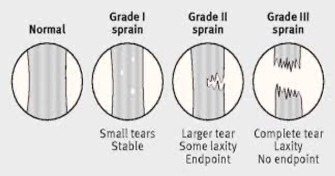
Injury classification typically uses a one to three scale, and denotes the severity and extent of ligament fiber disruption.
Grade 1 sprains will be locally tender and may include little or no noticeable swelling (effusion). Function will be impaired, but the athlete may be able to participate in training activities in an abbreviated or adapted fashion. Rarely, co-morbidities such as fracture, are associated. The ATF ligament and occasionally the CF ligament are involved. Crutches or immobilization are usually not required.
Grade 2 sprains will generally be acutely tender, swollen, discolored and carry an elevated risk of associated bone and or tendonous injury. Weightbearing tolerance will be compromised for days or weeks based on severity. Routine training will be correspondingly interrupted. The ATF and CF ligaments are typically injured. The Peroneal muscle tendon complex usually sustains a stretch injury and an “avulsion fracture” of the lateral malleolus (separation of a small bone chip) is not uncommon.
X-rays, crutches for a few days, activity modification and immobilization are important to shorten the overall time to symptom resolution. Unfortunately, because the injury is not agonizingly painful or completely limiting in terms of basic mobility, the athlete and caregiver accelerate the progression back to sport too quickly, only to regret that decision down the road. Resultant synovitis, excessive scar formation, capsulitis and anterolateral impingement are possible sequelae if management short cuts are taken. This may necessitate more complex future interventions (Jepson K, Solari J. Orthro Phys Ther Clinics of North Amer. 1994).
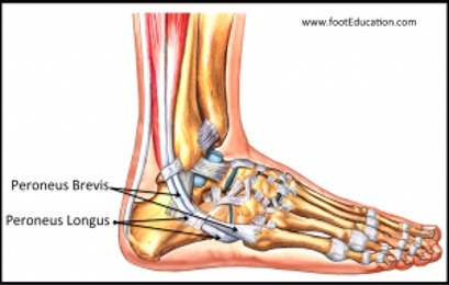
The Peroneal muscles are essential to dynamic stabilization of the ankle following a sprain. If inhibited due to stretch injury, they will be unable to contract with sufficient force to evert and dorsiflex the ankle (position of stability) while training. Restoration of Peroneal, and Anterior Tibialis muscle strength, endurance and speed of contraction is paramount to a successful outcome.
The Peroneal muscles are essential to dynamic stabilization of the ankle following a sprain. If inhibited due to stretch injury, they will be unable to contract with sufficient force to evert and dorsiflex the ankle (position of stability) while training. Restoration of Peroneal, and Anterior Tibialis muscle strength, endurance and speed of contraction is paramount to a successful outcome.
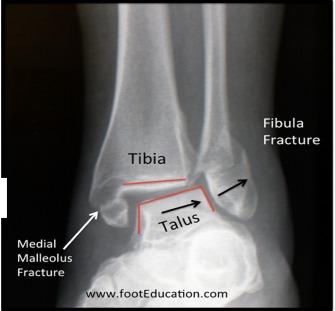
Grade 3 injuries involve complete disruption of the fibers, are tender, swollen. discolored, involve multiple ligaments and often have an associated fracture of the malleolar regions or proximal fibular shaft. There is no question in the athlete`s mind that something significant happened to the ankle. Complete and/or partial multi- ligament damage occurs including the ATF, CF, PTF and perhaps even the tissue connecting the distal tibia to the fibula (the interosseous membrane and Tib-Fib ligaments). The order of ligament injury occurs as listed above. Compromise of the ATF followed by CF damage, if the external forces persist and so on. If the distal “Tib-Fib” complex is injured, the term “high ankle sprain” is used. High ankle sprains are a different animal, often requiring 12 weeks or more to resolve, and typically require formal outpatient rehabilitation and occasionally surgery.
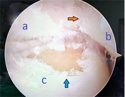
With grade 3 sprains, the talar dome articular cartilage or, “Teflon coating” of the joint surface, can be injured via compression and shearing forces. If mismanaged, internal joint injury can lead to long term problems such as post traumatic arthritis. An MRI or CT scan may be required to diagnosis such an injury.
Talar Dome articular surface damage below.
If forces are sufficient during injury, ankle dislocation can occur. This is a time sensitive injury as it can include vascular or neurologic compromise. Obvious deformity, open fractures, anterolateral foot sensory changes, “blanched” toe nail beds and a complete inability weight bear on the limb, are all “red flags”. Such injuries demand appropriate first aide, immediate ER evaluation and treatment by a trauma specialist.
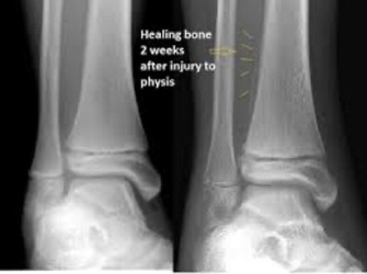
In young athletes, the possibility of Physeal (growth plate) injuries should not be discounted. Such injuries, classified as Salter-Harris Fractures, are not uncommon with grade 2 and above injuries in children. If you coach young athletes, err on the side of caution.
Type 2 Salter-Harris injury below.
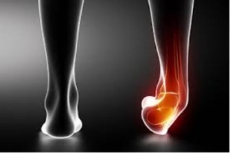
Significant Grade 3 ankle injury below.
Management of lateral ankle sprains involves a step process as with any orthopedic dysfunction. Rehabilitation concepts which are specific to Grade 2 sprains are a good “middle of the road” place to start, and will be outlined here. Grade 1 sprains will go through the same processes at an accelerated rate. Grade 3 injuries will likely require activity and timeframe adaptions due to “healing constraints” of secondarily injured tissues, such as bone.
Phase progression is based on competent completion of, and tolerance for, lesser demanding rehabilitation tasks. Time frames do not dictate a return sport. Not all Grade 2 sprains take “6 weeks” to resolve. Some progress faster, some slower.
As with other joint ligament injuries, the first trauma is typically the worst symptomatically and require strict adherence to rehab protocols. Recurrent injuries are less symptomatic, as the ligament tissue has been previously compromised, has “scared” in an elongated state and has lesser stabilizing capacity. The adverse result of “static” stability compromise, makes “dynamic” stability much more vital to a successful outcome. Dynamic stabilization is a long-term commitment. Ligaments, once elongated, do not return to their prior length.
Following diagnosis and injury classification, the goals of ankle sprain rehabilitation are;
- modulate pain and swelling
- maximize the vascularity and integrity of healing structures
- restore functional motion
- re-train and strengthen inhibited musculature
- enhance balance, agility and reaction time
- functionally return to sport
All of these criteria must be met prior to returning the athlete to unrestricted training or competition.
Let`s turn these goals into rehabilitation phases using a Grade 2 injury as a reference point. It is important to recognize that the phases blend with each. Components of a previous phase continue into the next.
Phase 1, initial healing, 0-2+ weeks.
This time frame starts immediate post injury and continues to week 2 +/-. Injury to the ligament fibers sets off a cascade of physiologic events. Cell wall disruption signals an influx of inflammatory cells, enzymes, growth factors and protein building blocks for collagen repair. Vascular bed repair and proliferation is vital to this process. Anything we can do to promote, and more importantly, not disrupt this initial phase of healing will help save time. Using crutches for a few days will help negate adverse mechanical input to the traumatized tissue. A 25-50% partial weight bearing technique should be employed with an emphasis on heel strike as tolerated.
Swelling and inflammation is a necessary evil. It is the process utilized by the body to mobilize building blocks to the injured region. The literature is unclear as to how much to suppress inflammation via medicines, ice, contrast, compression, etc. Please review a prior post on this site entitled; “Icing after injury; evidence based or folklore?”, to gain an appreciation of the debate. In general, anything that enhances circulatory exchange and normalizes the tissue environment will promote healing.
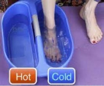
Pain management is important for the introduction of therapeutic exercise. Excessive effusion (swelling) will limit motion via distention pressures within the joint if allowed to persist. Occasional icing for 10-12 minutes via packs or submersion, along with compression, will help with introductory rehab activities tolerance and is unlikely to significantly interfere with the bodies healing response. Contrast baths have been shown to be effective regarding mobility gains. Compression is best achieved via an orthotic device, as “wrapping” with an elastic bandage application has been shown to be highly variable in terms of pressure and potentially harmful (Duffley H, et al. Ath Train. 1989).
Anti-inflammatory medicines should be used sparingly. Even over the counter varieties are quite powerful in terms of inflammatory suppression. Get the opinion of your physician or pharmacist. There is ongoing debate in the literature regarding the use of NSAIDs, particularly in the < 2 week post injury phase. Robust human studies are lacking (Schug S. Do NSAIDs Really Interfere with Healing after Surgery? J Clin Med. 2021, 10, 2359).
Elevation with active motion (ABC`s with the big toe), pneumatic garments, retrograde massage and gentle manual soft tissue release/mobilization techniques may be useful.
Electrical stimulation and ultrasound have been shown via animal studies to stimulate collagen production and vascular activity (Ramirez A, et al 1997. Frieder S, et al 1988. Jengyu L, et al 2007. Tsai WC, et al 2011).
The key with this phase is to recognize that the bodies response to injury serves a purpose and the athlete should respect that.
Phase 2, motion, 1-2+ weeks.
Range of motion activities should begin as soon as the athlete feels able. This phase will overlap with Phase 1. Motion activities will be utilized up to, and beyond the return of normal function. Pain levels exacerbated by motion exercise should be no more than 2 grades above the resting pain level on a subjective 0/10 scale. Discomfort should return to prior resting levels within seconds of exercise completion. If not, the load was too high. This concept will hold true for future therapeutic exercise.
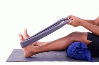
Non weight bearing (open kinetic chain) and partial weight bearing (closed kinetic chain) should dominant the exercise list. Extremes of available motion should be avoided as individual ligament bands come under greater tension at end ranges of the joint. Movement into dorsiflexion and eversion should begin first, while plantar flexion and inversion movements (the likely injury mechanism) should be added days later. Calf stretching with knee flexion will help negate the chance of joint motion restrictions due to tissue scaring, while not “fighting” against probable gastrocnemius tone. Four reps of 30-90 second holds are suggested, twice a day. For a skier, accessing adequate dorsiflexion is essential for technique, speed, balance, and stability. The ability to start DP work with competence early in the rehab process, is dependent on re-establishing knee and ankle motion (Holmberg HC, et al. Contribution of the Legs to Double-Poling Performance in Elite Cross-Country Skiers. Med Sci Sport Exer. 2006).
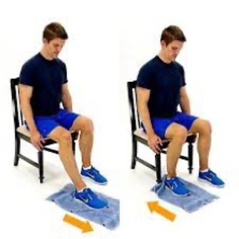
Heel slides sitting in a chair allow for early closed chain input with appropriate loading. A towel or sock lessens friction. The pain scale analog can direct extent of excursion. Four sets of 12-15 reps, is a good starting point. Side to side forefoot motions can be added when comfortable. Closing the eyes will introduce a proprioceptive component, vital to post sprain stability.
Intrinsic muscles of the foot, important for balance feedback and stability can be worked in the same position via picking up and dropping a small stone or marble in bare feet.
Stationary cycling with a high seat and low tension is usually tolerated within a couple of days post injury and beneficial for vascular bed proliferation and fluid exchange. Aquatic based exercise is appropriate in the absence of bony involvement. Fins should not be used, as lever system and torque inputs should be avoided at this juncture.
An ergometer may be used provided the affected side is shod and wrapped. Normal duration should be abbreviated. Core exercise on a mat is appropriate. No plank postures that load the foot and ankle should be utilized however. Upper body gym work can continue if the athlete is seated.
NO FINS!
Walking should be limited to household duties only. Driving may be appropriate based on right or left ankle involvement, transmission, etc.
Phase 3, re-activation and strength, 2-4+ weeks
With a grade 2 lateral sprain and above, it is very likely the individual also sustained a concurrent Peroneal musculotendinous strain injury. Excessive inverted positioning, required to disrupt the ATF and CF fibers, also places significant load on the lateral compartment musculature. As primary evertors of the ankle/foot complex, the Peroneal Longus and Brevis reflexively contract vigorously to dampen potentially injurious inversion.
If external forces exceed the tensile characteristics of the Peroneals, a strain of those fibers ensues. “It`s just a strain,…” , posted on this site last summer will provide some insight.
Highly functioning peroneal muscles are essential to ankle stabilization post injury. They help prevent recurrent sprains and symptoms, which are documented to be as high as 73% as noted previously. Any muscle tendon unit experiencing irritation directly, or via reaction to adjacent joint damage, responds by shutting down. This is termed “Arthrogenic Muscle Inhibition” (Rice DA, McNair PJ. Semin Arthritis Rheum. 40,2010). In short, injury causes weakness via neurogenic inhibition which requires “focused activation” to regain full strength and power. A gradual return to one`s sport alone does not meet the criteria of “focused activation”.
Strength exercise progression in phase 3 consists of; sustained isometric, to eccentric, to concentric, to functional closed chain, to plyometric, and finally sports specific training.
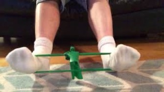
Sustained “light” isometrics provide a cellular regenerative stimulus without causing further damage. Sixty to 90 second hold times at a mid- range joint position is suggested, 4 reps daily, twice if tolerated well. Band, manual or fixed object resistance can be employed. Isometrics can begin when they meet the “2 grades over resting pain” criteria mentioned previously. Isometrics should continue for 2 weeks on a daily basis and should be utilized periodically (weekly) long term, to ensure healthy tissue composition.
Evertor iso`s below.
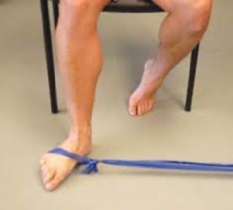
Eccentric exercise refers to the controlled elongation of a muscle under load. “Cheat up, down slow” is a common clinician cue for technique description. The controlled lowering/resisting portion should take 4-6 seconds and the repositioning of the weighted ankle/foot to the starting point may require an assistant to lift the body part into position. An option for solo use, would be to utilize elastic resistance. Move the joint into the starting position, then stretch the band and “fight” the load. Four sets of 4 reps at moderate resistance, 4 x/week for 2-3 weeks is suggested. The athlete must recover completely between sessions. Early focus is on the evertor and dorsiflexor groups.
Cheat out, back slow in,……..
Concentric work follows a typical rep and set design with an emphasis on endurance work via multiple sets of 15-20 reps. Free weight, bands, manual, or machine-based resistance sources may be used provided pain tolerance criteria is met and no residual swelling ensues. On-line programs are plentiful.
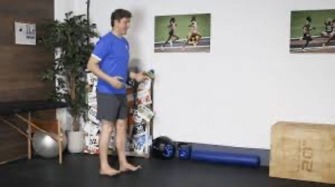
Closed chain activities include squats, lunges, leg presses, lateral walks, cycling intervals, range limited calf raises, etc. Body weight only, progressing to added dumbbell weight is suggested. Heel walking for time (15-60 seconds) x 4-6 reps, is a great way to work the endurance component of the dorsiflexors and evertors so vital for ankle stability and ski retrieval under one`s center of mass. Use of shoes is suggested to avoid bruising of the calcaneal fat pad.
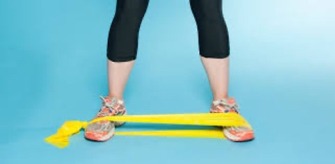
Lateral band walks with resistance attached high (knees) and low (forefoot) work the lateral column, hip down, in its entirety. In a 2010 study by Rahnama et al, subjects with functional ankle instability demonstrated poor postural hip and core control which was further compromised when a cognitive task was introduced (Attentional Demands and Postural Control in Athletes With and Without Functional Ankle Instability, J Ortho Sports Phys Ther 40 (3)). Not great for roller skiing in traffic!
Phase 4, balance, agility and proprioceptive 3-6+ weeks.
Kinematic work options are limited only by the athlete`s imagination. All prior strength work can simply be performed on uneven or unstable surfaces. Bosu balls, wobble pads, pillows, 1x3`s, sand, etc, all provide added neuromuscular challenge. Change of direction walking drills upon cue, pattern walking (ie zig zag) and agility courses involving step overs, heel toe walking, and slide boards for skating work all hone balance, reaction time and dynamic stabilization. Arguably this is the most vital of progressions for athletes who experience numerous sprains throughout their lives. Chronic ankle instability and balance deficits are well correlated (Brown and Mynark, J Ath Train. 42(3); 2007). Injecting balance challenges weekly into one`s normal training plans is paramount to reducing recurrent injury.
Return to running progressions are prevalent. This site offers excellent protocols. If this phase of rehab goes poorly due to anterolateral pain, plateaued motion/dorsiflexion or swelling, the athlete is encouraged to seek out clinical evaluation. “Impingement Syndrome” of the ATF region is not uncommon. It is resultant of excessive anterolateral capsular scarring and requires specific mobilization techniques best administered by a qualified Physical Therapist (SCS or OCS). A PT can also make recommendations for appropriate diagnostic studies.
Phase 5, functional return to sport, 5+ weeks
This phase should be prefaced with functional movement, balance and strength testing. A passing grade is simply a return to symmetry. “It feels great”, is not a passing grade. Seek out a qualified clinician to perform a battery of tests.
Returning to full training (snow or road) involves double pole work first, followed by “drills and skills”, skating and finally classic skiing, perhaps in combi boots to begin. Any OD sessions should be done on a bike. Bounding and speed workouts will be introduced last in the progression.
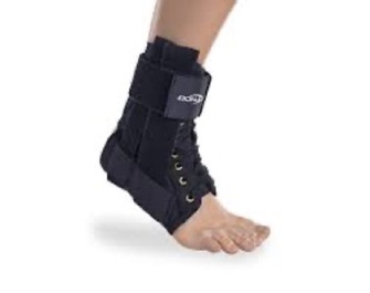
There is no down side to ankle orthosis use, provided it fits in shoe wear and does not abrade the skin. Ankle taping is not recommended as the adhesive loosens with perspiration and the fabric stretches when saturated. Ankle “braces” are more stretch resistant than tape and can be “laced” tighter mid- session. They provide superior support, may enhance the neuromuscular feedback system and do not create “reliance” (Rovere G.et al, Amer J Sport Med).
Compliance with an appropriate rehabilitation program will eliminate the need for long term bracing.
Lateral ankle sprains are prevalent in the athletic population. Due to their commonality, they are frequently under diagnosed and mismanaged. Even low-grade injuries can plague an athlete for months or years. Do it right the first time. You won`t be sorry.