
ANTERIOR HIP PAIN
By Kurt Jepson
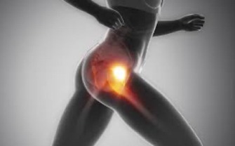
In addition to structural joint loads associated with flexed positions, dramatic contractile forces emanating from the core and hip musculature add to hip joint stress. As the athlete moves into the pre-comp phase of training, exposure is amplified. Hips therefore, are predisposed to overuse or acute injury. Frontal or anterior hip pain is a common problem with dynamic athletes.
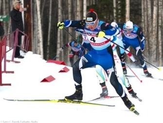
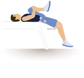
As skiers, we spend a vast amount of our training time engaged in activities which include, in fact are mandated from a technique standpoint, the use of flexed hip and core postures. Cycling, skiing, ice skating, bounding and hiking all fall into this “flexed posture” category.
In addition to structural joint loads associated with flexed positions, dramatic contractile forces emanating from the core and hip musculature add to hip joint stress. As the athlete moves into the pre-comp phase of training, exposure is amplified. Hips therefore, are predisposed to overuse or acute injury. Frontal or anterior hip pain is a common problem with dynamic athletes.
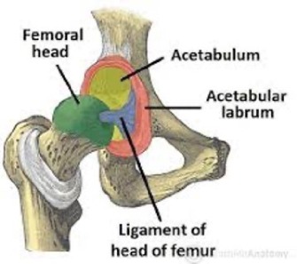
Symptomatically, anterior hip issues present with consistent or transient pain, and often “snapping” due to inflamed tissue subluxation. This can be alarming, as the athlete has a sense that the hip joint is unstable or damaged. The issue however is typically external to the joint proper and does not involve articular (internal) anatomy. Exceptions include significant arthritis (in older athletes) or labral pathology. Dysfunction of the cartilage rim of the hip (Labrum) is typically resultant of traumatic input, congenital anatomic anomalies and/or extreme training loads.
Differentially, osteoarthritis (OA) tends to “ache” at rest, feel better with low load activity (ie cycling) but worsen as the activity intensifies or progresses. Running rarely goes well. Symptoms persist post activity and may be bilateral. Range of motion loss is pathopneumonic and the patient will find tasks such as tying shoes and crossing one`s leg to don socks increasing challenging as time progresses. Anti-inflammatory meds and rest days typically lead to improvement and can be somewhat diagnostic if a trend is recognized. Past trauma to the hip can escalate OA in patients that would typically not fit the profile chronologically.
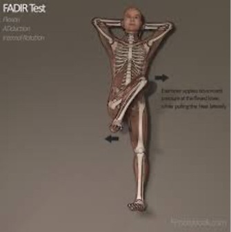
Labral issues are often immediately reactive to abrupt loading forces (ie running down inclines, deep squats) and are provoked via the hip being fully flexed, adducted and internally rotated. This is referred to as the Impingement Sign of the Hip (Hananouchi T et al, Anterior Impingement Test for Labral Lesions Has High Positive Predictive Value, Clin Ortho Relat Res 2012, 470: 3524-3529). Labral injuries are typically unilateral. Management is complex and beyond the scope of this post.
Stress fractures, circulatory problems, cystic activity, and in young skiers, growth plate issues, are rare but do happen.
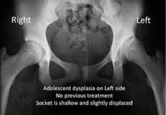
If the athlete suspects one of these etiologies and has an associated history, a trip to your sports med physician for base line films is warranted. X-rays are also important to differentially rule out remote pathologies which refer pain to the hip (ie low back) or identify architectural anomalies such as dysplasia.
In endurance athletes, anterior hip pain is typically soft tissue based. The specific origin of the problem can be clinically challenging as numerous regional anatomic structures can be at fault.
A work by Hammound S, et al published in Sports Health, Mar-April 2014, takes a deep to superficial “layered” approach to understanding functional hip anatomy. The authors describe layer 1 as being comprised of the bony and cartilage components, layer 2 the enveloping ligamentous capsule and bursae, layer 3 the surrounding musculotendinous groups and layer 4 the neurokinetic components. Most non-traumatic/overuse symptoms arise from layer 2 or 3.
Layer 1 components.
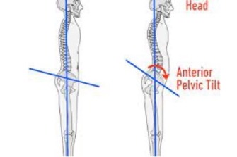
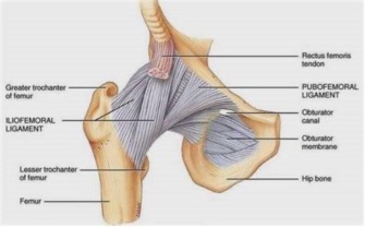
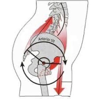
As with the shoulder joint, ligamentous irritations about the hip result from mobility imbalances of the joint capsule. Commonly, Nordic skiers have anterior capsular restrictions and posterior laxity. This is the result of years of training induced anterior pelvic tilt (“Dumped Pelvis”), hip flexor over-development and perhaps gluteal weakness. Prior trauma may also predispose the capsule to anterior/posterior tension imbalance resultant of scar deposition and/or attenuation
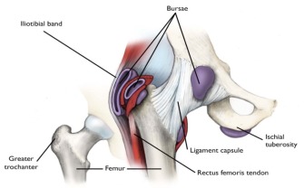
These factors can lead to poor tissue interface separation between layer 1 and 2. The result is frictional irritation at the trochanteric and/or acetabular rim (socket) regions as the hip moves from flexion/ internal rotation toward extension/external rotation (ie stepping up the hill in V1) as ligament tissue translates over these prominences. This can manifest as frontal hip pain and “snapping”. These symptoms typically start gradually and worsen in line with training duration. Over time the frictional irritation can induce scar tissue formation or even calcification, which of course exacerbates symptoms. Bursal “sacks” lying between layers are frequently involved. Bursal tissue is highly innervated and quite painful when irritated.
Layer 2, ligamentous capsule
Restrictions in layer 2 can limit V1 motion and efficiency.
Poor layer 2 (capsular-ligamentous) mobility increases the metabolic cost of movement and also places other structures under stress. When functional motion demands exceed what is available at the hip, the hemi-pelvis and lumbar spine are forced to contribute motion and thus subject to injury.
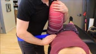
Layer 2 “hypomobility” is best addressed by a physical therapist skilled at “targeted” joint mobilization and distraction techniques. A capsule is rarely uniformly restricted and identifying the specific limitation requiring mobility enhancement is important. Abrupt manipulation techniques and aggressive stretching are counterproductive as the resultant micro-trauma to the capsule will only stimulate further scarring. Capsular tissue requires long sustained tensile input below “rupture” force levels to plastically deform and gain length.
Layer 2 “hypermobility” also increases metabolic cost of movement via different demands. The need for excessive hip muscular activity to dynamically stabilize the joint in the absence of adequate static ligament support elevates energy expenditure.
Anterior to posterior capsular tension must be balanced to ensure sound arthrokinematics and efficiency.
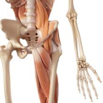
Layer 3 anatomy includes the surrounding musculotendinous groups. This includes those that insert locally and those that cross the hip as two joints muscle units. Two joint muscles have numerous core and hip joint functional responsibilities and are therefore more exposed to injury and overuse (Neumann D. Kinesiology of the Hip: A Focus on Muscular Actions. JOSPT 2010; 40(2)). The Iliopsoas, adductors and Rectus Femoris are most notable.
A review of the April 2020 post on this NTS site entitled “Hip Flexors and Athletic Posturing” will assist the reader in understanding the anatomy and functional role of the Iliopsoas and Rectus Femoris as core influencers and prime movers.
Two joint hip muscles.
Deep Rotators.
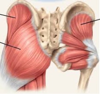
The deep rotators, similar to the rotator cuff of the shoulder, act as “dynamic ligaments” guiding proper roll and glide of the ball and socket assembly while providing compression and stability. Their function is vital to the hip. When untrained and/or under-activated, larger muscles of the hip must compensate for them in addition to their primary responsibilities of power generation.
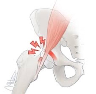
The Iliopsoas is an example of a “universal compensator”. The “psoas” is anatomically unique in that regardless of the relative flexed or extended position of the hip, it can deliver significant contractile load to move or stabilize the hip joint. It is a strong internal and external rotator based on joint position. The psoas is not subject to relative positional weakness as most muscles are. It is very active during most abdominal tasks (Juker D., McGill S. et al Med Sci Sports Exer; 1998). It gets very little rest. In the absence of sufficient deep rotator strength, it often falls victim to overuse irritation (Rajkumar RV. Force couple mechanics on the femur during closed chain activities of the lower limbs. Int J Physiother Res.2014; 2(6): 766-771).
Iliopsoas tendinitis and insertional strains are common overuse problems in endurance athletes. Often these injuries are diagnosed as a simple “groin or hip flexor pull”. If inadequately addressed, long term involvement leads to; low back pain, “snapping/popping”, secondary bursitis, flexor weakness and extension deficits of the hip joint.
These issues feed into the afore mentioned “dumped pelvis” scenario and limit efficient skiing due to posterior column and core inhibition.
Unfortunately, many treatment protocols address psoas irritations with a program consisting of; stretching, ice, massage, anti-inflammatory modalities and activity modification. The root cause of psoas irritation must be addressed. Deep rotator, as well as gluteal, weakness must be corrected to offload the psoas as the athlete resumes training. Exercises must be selected that adequately recruit rotators and gluteal muscles, without placing undesired stress on an already overworked psoas complex. Philippon et al published an excellent work on this topic in 2011 (Rehabilitation exercise for the gluteus medius muscle with consideration for iliopsoas tendinitis. Am J Sports Med. 2011; 39(8)).
Stretching in the acute phase of psoas injury is contraindicated. A strained hip flexor will not do well with added tensile strain beyond its current physiologic limits. Targeted flexibility work should be delayed until symptoms resolve. Stretching also does nothing to address underlying weakness.
Rotator strengthening should be initiated with sustained, submaximal isometrics (60-90 sec x 4 reps) progressing to eccentric, to concentric, to functional multiplanar movements and lastly a plyometric progression. This progression is similar to that utilized for any muscle tendon dysfunction (see the May 2020 Nordic Team Solutions post on Eccentrics). Pain latency parameters should be honored as described in this NTS article.
In addition to the deep rotators of the hip, the Gluteal musculature must be fully rehabilitated. The superior aspect of the gluteus maximus in particular has important rotational control responsibilities, most notably when the hip is in flexed postures as in skiing (Delp SL et al. J Biomech. 1999, Selkowitz DM et al. JOSPT; 2016). It is important to include multiplanar strengthening motions for the hip. Resistive hip extension with combined external rotation (toe out) movements will train the “glut max” to be a powerful extensor and a concurrent rotational stabilizer of the joint. All selected exercises should be completed in a “snap free” range of motion. There may be a small motion window in the beginning but that is fine. Any crepitus/popping/snapping should be avoided during rehab, as frictional inputs from a poorly controlled hip joint will elevate symptoms.
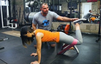
Quadruped band hip extensions with external foot position/”out toe”.
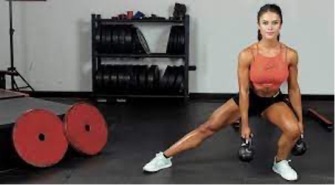
Kettlebell lateral lunge eccentric down to concentric “quick” up with “out toe”.
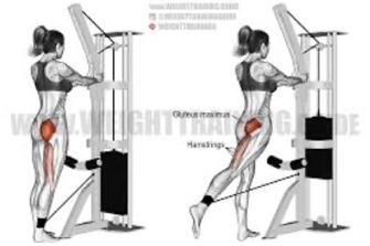
Standing cable hip extension with simultaneous external rotation.
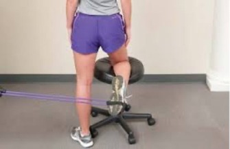
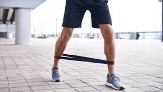
Retro oblique band walk emphasizing “out toe”.
Utilize any of your favorite hip strengthening exercises and add external rotation to the movement. Be as inventive as you wish with the goal of engaging various muscle groups in multiple planes while extending and rotationally stabilizing the hip joint.
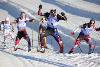
Sounds a bit like skiing…..
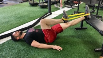
As strength gains are realized and rotator control improves, the frictional irritation will lessen anteriorly and “snapping” if present, begins to subside. At this point, program additions focus on building durability of the iliopsoas and rectus femoris musculo-tendonous regions. Sustained isolated hip flexor isometrics (60-90 sec x4) in various positions are initiated and the gradual addition of eccentric resistive exercises (sets of 6 reps) are included as tolerated.
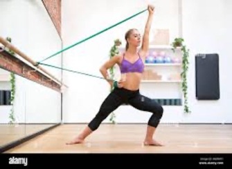
Combined hip flexion band iso on right, with concurrent left extensor recruitment.
The last phase of layer 3 rehabilitation includes hip flexor and quadriceps stretching, Yoga, foam rolling, and a logical return to skiing. This where the athlete should commit long term to minimizing anterior pelvic tilt via gym work and reinforce posterior pelvic tilt while skiing.
It might appear that management of anterior hip pain is counterintuitive, specifically, work the back to fix the front! That is indeed what it takes to resolve layer 2 and 3 issues, resulting in a favorable outcome.
Layer 4 issues are perhaps less frequent. Superficial contusions, skin lesions, peripheral sensory nerve pathology and myofascial dysfunctions can all have a negative effect on proper motor function about the hip as well as balance. Refer to a post on this site from December 2020 entitled “Balance 101” to get a feel for neurokinetic management strategies.
Anterior hip pain in Nordic athletes can be a stubborn and reoccurring issue due to adaptive, technique mandated postural factors. Successful management focuses on correctly identifying the offending “layer”, correctly assessing tissue dysfunction and employing targeted treatment interventions to negate imbalance and deficiencies.